
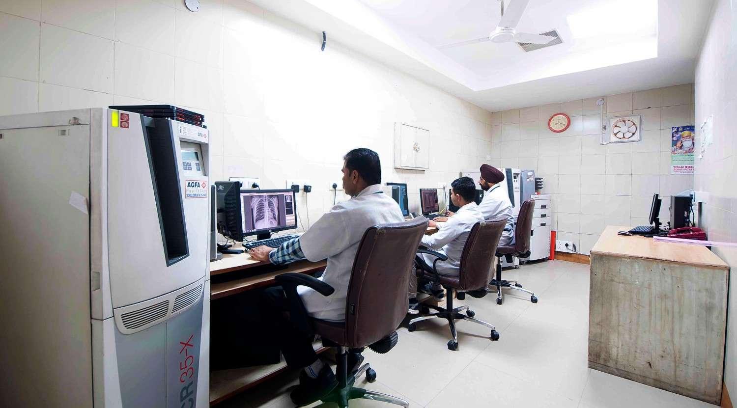
The Department of Radiology at Dayanand Medical College, Ludhiana, is a center of excellence equipped with state-of-the-art technology to deliver precise and reliable diagnostic imaging. Our services encompass a wide range of advanced imaging modalities, supported by a team of highly experienced radiologists and technologists committed to patient care and safety.
Our Services and Specializations
Digital X-rays
- Fully digital imaging system for high-resolution images with reduced radiation exposure.
- Comprehensive diagnostic solutions, including specialized imaging for trauma, orthopedic, and chest studies.
Advanced Ultrasonography
- Elastography: For assessment of liver fibrosis, breast, and thyroid abnormalities.
- Intestinal Ultrasonography: Specialized imaging for bowel disorders such as IBD.
- Fetal Ultrasonography: High-resolution imaging for monitoring fetal development and anomalies.
- Doppler Imaging: Evaluation of blood flow in vessels and assessment of vascular abnormalities.
- Musculoskeletal Ultrasonography (MSK): Detailed imaging of joints, tendons, and ligaments.
- AV Fistula Doppler: Critical evaluation of arteriovenous fistulas in dialysis patients.
Barium and Contrast Investigations
Comprehensive gastrointestinal imaging, including:
- Barium Swallow and Barium Meal: For esophageal and stomach disorders.
- Barium Enema: For detailed imaging of the colon and rectum.
Advanced contrast-based studies, including:
- Hysterosalpingography (HSG): Evaluation of the uterus and fallopian tubes for infertility investigations.
- Intravenous Pyelography (IVP): Detailed imaging of the urinary system.
- Retrograde Urethrogram (RGU) & Micturating Cystourethrogram (MCU): Assessment of urinary tract abnormalities.
CT Imaging
Somatom Siemens Force CT scanner (384-slice):
- Dual-source technology for ultra-fast imaging with low-dose radiation.
- Special applications: Cardiac and coronary CT, virtual colonoscopy, detailed vascular studies.
Emergency CT (Siemens Somatom Go Up, 64-slice):
- Dedicated CT system for rapid and critical diagnostic needs.
MRI Services
3-tesla Siemens Magnetom MRI machine:
- Advanced applications, including:
- Neuroimaging: Tumors, stroke, and epilepsy diagnostics.
- Cardiac MRI: Structural and functional cardiac imaging.
- MR Enterography: Imaging of the small intestine for conditions like Crohn’s disease.
- Neurography: High-resolution imaging of peripheral nerves.
- Dynamic Angiography and Perfusion: Precise vascular evaluation.
- Fetal MRI: In-depth assessment of fetal anatomy and anomalies.
Mammography
Digital Mammography: High-quality breast imaging for early detection of breast cancer and other abnormalities.
DEXA (Dual-Energy X-ray Absorptiometry)
- Equipped with the advanced Horizon Wi DEXA machine for precise bone health assessments and body composition analysis, offering superior diagnostic accuracy and the ability to diagnose osteoporosis effectively.
- Gold-standard imaging for bone mineral density (BMD) assessment to diagnose osteoporosis and monitor bone health.
State-of-the-Art PACS (Picture Archiving and Communication System)
- Advanced PACS technology for seamless image storage, retrieval, and sharing.
- Ensures secure, real-time access to diagnostic images across departments for efficient and integrated patient care.
Additional Facilities
Portable X-ray and Ultrasonography
Provision for portable X-ray and ultrasonography to cater to the diagnostic needs of critically ill and moribund ICU patients, ensuring timely and accurate imaging at the bedside.
Horizon Wi DEXA Machine
Equipped with the advanced Horizon Wi DEXA machine for precise bone health assessments and body composition analysis, offering superior diagnostic accuracy.
Why Choose Us?
- Cutting-Edge Technology: Equipped with the latest imaging equipment for accurate diagnostics.
- Expert Team: Experienced radiologists and technologists dedicated to quality and patient safety.
- Patient-Centric Care: Comfortable and safe imaging procedures tailored to individual needs.
- Emergency Services: 24/7 availability for rapid diagnostics in critical cases.
Interventional Radiology (IR) Unit
The Interventional Radiology (IR) unit DMC&H, Ludhiana, stands at the forefront of modern minimally invasive medical care. It is one of the leading centers of interventional excellence in North India, offering a wide spectrum of image-guided diagnostic and therapeutic procedures that combine precision, safety, and innovation.
Equipped with the latest high-end fluoroscopy systems, advanced Digital Subtraction Angiography (DSA) suites, CT and MRI guidance, and state-of-the-art ultrasound platforms including Contrast-Enhanced Ultrasound (CEUS), the department provides world-class interventional services for patients requiring complex yet minimally invasive care.
By integrating cutting-edge imaging with skilled expertise, the department enables patients to benefit from treatments that are less painful, less invasive, and more effective than traditional surgery.
Vision and Mission
Our philosophy is grounded in the belief that: “Precision in Imaging is Precision in Healing.”
We aim to provide comprehensive, compassionate, and technologically advanced image-guided care that reduces morbidity, enhances recovery, and maintains the highest global standards of safety and efficiency.
Our Mission Includes
- Delivering state-of-the-art interventional radiology services across all organ systems
- Promoting research, innovation, and continuous learning through academic collaboration
- Offering structured training programs for residents to nurture future IR specialists.
Key Advantages of Interventional Radiology
- Minimally invasive procedures with no major surgical incision
- Performed under local anesthesia or conscious sedation
- Less pain and complication rates
- Shorter hospital stay and early return to routine life.
- Cost-effective compared to conventional surgery
- Highly cost effective compared to surgery
Technology and Infrastructure
The department is equipped with the most modern, globally benchmarked interventional radiology infrastructure to ensure diagnostic accuracy and procedural excellence.
Fluoroscopy and Angiography
- Advanced flat-panel Digital Subtraction Angiography (DSA) system with 3D rotational angiography, Real-time road-mapping and radiation dose-reduction technology
CT- Guided Interventions
- CT-guided procedures for deep or complex lesions
- Dedicated hybrid interventional suite with sterile OT setup for vascular and non-vascular procedures
Ultrasound and CEUS
- High-end ultrasound systems with multi-frequency linear, convex, and micro-convex probes
- Contrast-Enhanced Ultrasound (CEUS) for:
- Advanced lesion characterization
- Tumor vascularity evaluation
- Post-intervention assessment
- Targeted biopsies and interventions
- Portable ultrasound systems for bedside and ICU-based procedures
Support Facilities
- Advanced anesthesia services with continuous patient monitoring
- Dedicated post-procedure observation and recovery area
- Trained nursing and technical staff
- Strict adherence to radiation safety and infection control protocols
Range of Interventional Services
Vascular Interventions
- Diagnostic angiography
- Angioplasty and stenting of arteries and veins
- Dialysis access maintenance (AV fistula / graft)
- Permacath insertion (Dialysis catheter)
- PICC line insertion
Hepatobiliary intervention
- Transjugular Liver biopsy (TJLB)
- Transjugular intrahepatic portosystemic shunt (TIPSS) for refractory ascites and bleed in cirrhosis
- BRTO/PARTO for endoscopic refractory variceal bleed in cirrhosis
- Partial splenic artery embolization
Peripheral vascular Interventions:
- Varicose vein laser/ Glue ablation
- Peripheral arterial disease (PAD) interventions
- Deep vein thrombosis (DVT) thrombolysis and mechanical thrombectomy
- Pulmonary embolism interventions
- Sclerotherapy
Urointerventions:
- Varicocele embolization
- Prostatic artery embolization
- Antegrade DJ stenting
Thyroid intervention
- Thyroid nodule ablation and thyroid artery embolization
Non-Vascular Interventions
- Image-guided biopsies (liver, lung, bone, lymph node, soft tissue)
- Image-guided drainage of abscesses and fluid collections
- Percutaneous nephrostomy
- Biliary drainage and stenting
- Gastrostomy and jejunostomy tube placement
Oncology Interventions
- Tumor ablation techniques
- Microwave ablation (MWA)
- Radiofrequency ablation (RFA)
- Cryoablation
- Transarterial chemoembolization (TACE)
- Transarterial radioembolization (TARE)
- Palliative interventions for cancer-related pain and bleeding
Spine and Pain Management Procedures
- Vertebroplasty for painful osteoporotic or pathological vertebral compression fractures
- Kyphoplasty for vertebral body height restoration and pain relief
- Transforaminal steroid injection for radicular pain due to disc prolapse or spinal stenosis
- Genicular Artery embolization
- Celiac plexus block
- Lumber sympathectomy
Emergency and Trauma Interventions
- Emergency embolization for internal bleeding
- Gastrointestinal bleed embolization
- Post-traumatic vascular injury management
- Post-partum hemorrhage : Uterine artery embolization
- Hemoptysis : Bronchial/ Pulmonary artery embolization
Women’s Health Interventions
- Uterine fibroid embolization (UFE)
- Pelvic congestion syndrome embolization
- Management of obstetric and gynecological bleeding
Fetal intervention
- Amniocentasis
- Intrauterine blood transfusion
- Fetal reduction
- Radiofrequency ablation in Twin Twin Transfusion syndrome
Academic Activities and Training
- Regular academic teaching sessions for postgraduate residents
- Hands-on training in image-guided procedures
- Case discussions and morbidity-mortality meetings
- Participation in multicentric research projects
- Encouragement of publications in peer-reviewed national and international journals
- Active involvement in workshops, CMEs, and conferences
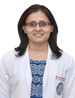
Head of Department
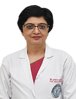
DR. KAVITA SAGGAR
Professor & Head


About Doctor
With decades of experience in radiodiagnosis, she has special interest in Neuro imaging and body imaging. She has held key academic positions and contributed significantly to medical research, with 60 national and 32 international publications. A fellow of the Indian College of Radiology & Imaging, she has served as President of the Punjab State Chapter of IRIA and held various leadership roles. She has also received multiple awards, including the Swabhimaan Award by Noble Foundation. Read More
Dayanand Medical College & Hospital

.webp)

.webp)
Enter at least 2 characters...
Enter at least 2 characters...
Enter at least 2 characters...
Enter at least 2 characters...
Enter at least 2 characters...